These radiographs show the progression of Taj's bone healing. Please click on any image for a close-up view.
Immediately post-break:
 |
| Lateral View: Post Break |
 |
| Front to Back View: Post Break |
|
Post-splinting (immediately prior to surgery):
 |
| Lateral View1 : Post Splint |
 |
Front to Back View:
Post Splint |
 |
| Lateral View 22: Post Splint |
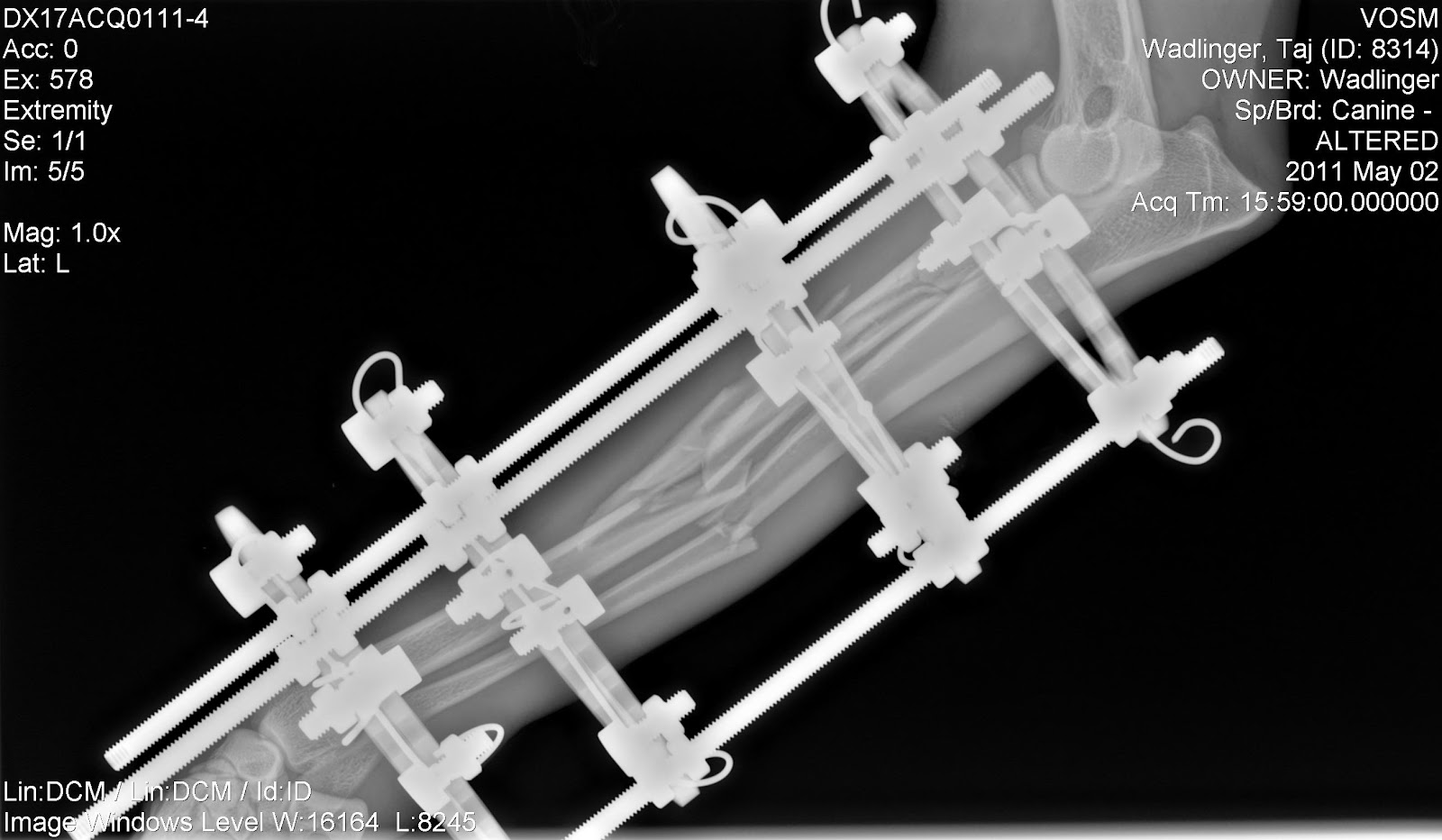
Post-surgery:
Post-operative radiographs confirmed appropriate implant positioning and good anatomic alignment (elbow over wrist). Perfect reduction was not attempted due to the severe comminuted nature of the fracture. Perfect positioning of the bone fragments is not necessary, and clinical studies have shown that disturbing a break with internal surgery can sometimes: slow the healing process by disturbing the fracture hematoma and periosteal blood supply, cause longer surgery times, heighten risk for infection and blood loss, cause higher levels of post-op pain, and possibly more post-surgical complications (Dudley et al.,
J Am Vet Med Assoc., 1997; Johnson et al,
J Am Vet Med Assoc., 1998; Ozsoy & Altunatmaz,
Vet Med Czech, 2003; Schmal et al.,
J Orthop Trauma, 2011).
 |
| Lateral View: Post Fixator Surgery |
 |
Front to Back View:
Post Fixator Surgery |
|
4-weeks post-surgery:
Although the healing is
slow, these radiographs show evidence of healing. If you look closely (click the images to zoom in), you can see the beginning of a cartilage callus formation between some of the fracture sites. For lack of appropriate terminology, this looks like"cloudiness" or a "cloudy blob" forming between the fracture sites. This means that the bones are starting to mend together. The bone alignment is still retained from fixator application - which is good! Over the course of the next month, the callous formation should grow and this should begin to better fill in the break sites and start to pull some of the fragments together. Although it is slow progress, this is still progress nonetheless!
 |
| Lateral View 1: 4-weeks |
 |
| Lateral View 2: 4-weeks |
 |
| Front to Back View: 4-weeks |
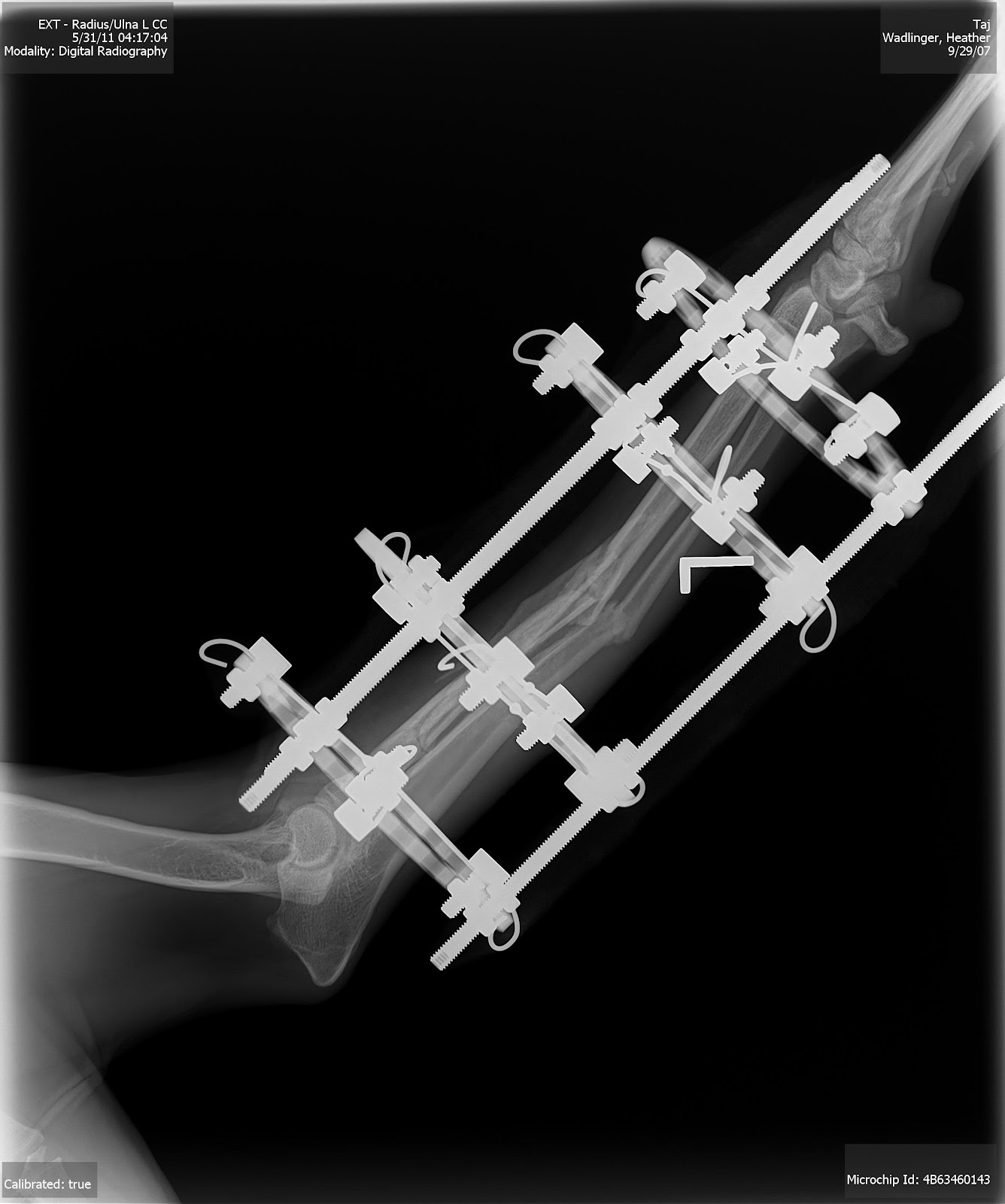
8-weeks post-surgery:
The cartilage callus is expanding! If you look closely (click images), you can see that the ulna is starting to re-adhere together as one continuous unit now! While the radius remains fractured in several pieces, there is
much more callus forming than in the 4-week radiographs. The bone alignment continues to be retained very well. I asked about that one shard of the radius that looks like it is "jutting out," but my vet assured me that if it does not end up being pulled back together with the callus formation over the course of the next two months that the body will naturally begin to reabsorb the unnecessary fragments (which boogles my mind). Over the next month, the radius should show more significant repair and any remaining gaps should fill with hyaline cartiledge and woven bone (weak bone made of a more haphazard organization of collagen fibers) - eventually transforming into lamellar (trabecular) bone (strong bone which has regular parallel alignment of collagen sheets). Over time and leg-use all this then hardens to compact (cortical) bone. This is good news - the healing is slow and steady - and by 12-weeks there should be a significant difference!
![]() |
| Lateral View: 8-weeks |
![]() |
| Front to Back View: 8-weeks |
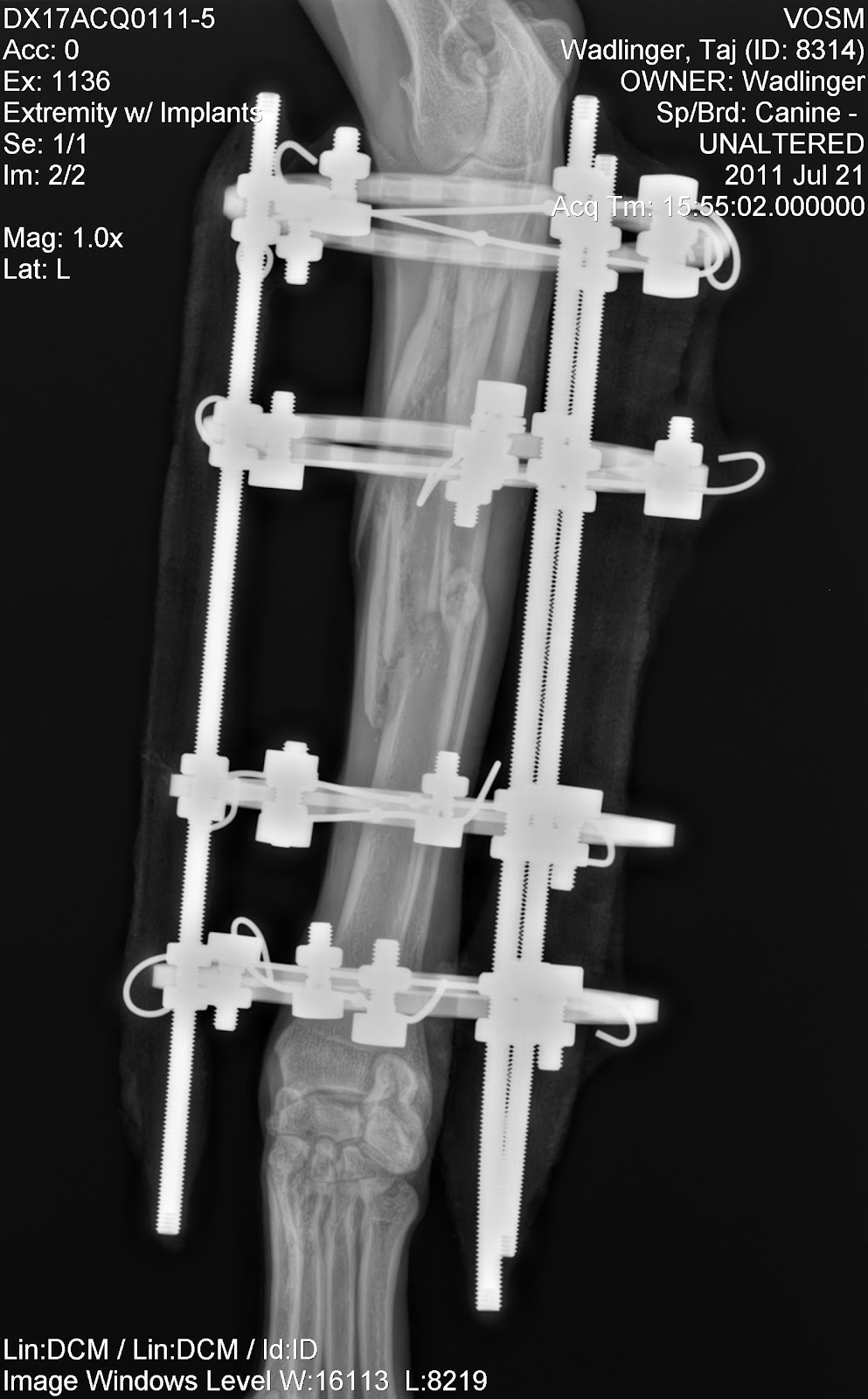
11.5-weeks post-surgery:
After starting the Exogen treatments, VOSM asked us to come down to Annapolis for the 12-week radiographs. There radiographs were taken after 10 ultrasound treatments. Dr. Christopher at VOSM gave us a thumbs-up and said that the progress from 8-weeks (in terms of callus formation) had significant improved. He believes we are still on track for a 16 week +/- fixator removal. You can see where the callus has filled in - especially in the "Front to Back" view (click on image below). Dr. Christopher wanted to see more callus in some of the more outlying radial areas before fixator removal can be considered, but we should be able to do another 30+ ultrasound treatments before the next radiograph visit! You can see in these x-rays that a lot of the hairline fractures have filled in quite nicely (scroll up to compare to the post-surgery fixator pictures - BIG difference!). There are still a couple areas (near tips of radial bone fragments) that need some more callus formation, but an overall positive visit!
 |
| Lateral View: 11.5-weeks |
 |
| Front to Back View: 11.5-weeks |
To come: Radiographs taken 16-weeks post-surgery...